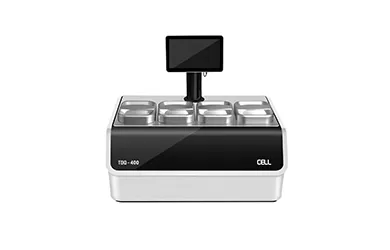
“Tissue Processing” refers to a series of necessary steps that prepare animal or human tissue from the fixation stage to the state of being sufficiently infiltrated with appropriate histological wax, allowing it to be sectioned on a microtome.
Laboratory managers often emphasize the importance of tissue processing to their staff. It is crucial to understand that using an inappropriate processing schedule or making fundamental errors (such as incorrect replenishment or sequencing of reagents) can result in tissue specimens being unsectionable. This means they will not yield any useful visual information. This situation is particularly severe in human diagnostic tissues because entire specimens are processed in a single batch. If the tissue is damaged in such cases, there may not be spare tissue available for further analysis, leading to a situation where the lab must explain to the patient why a diagnosis could not be made. Although mechanical or electrical failures of the automatic tissue processing machine can occur, most processing issues are caused by human errors. Therefore, emphasizing the importance of proper education and training for tissue processing personnel is crucial. Carefulness must be exercised when setting up processing programs for any run to ensure the best results.
1. Collect Fresh Samples
Fresh tissue samples come from various sources and are prone to damage when removed from patients or experimental animals. Therefore, they must be handled carefully and fixed as soon as possible after dissection. Ideally, fixation should occur at the site of extraction, such as in the operating room. If this is not feasible, it should be fixed immediately upon arrival at the laboratory.
2. Fixation
The specimen is immersed in a liquid fixative, such as a formaldehyde solution like formalin. This agent gradually infiltrates the tissue, inducing chemical and physical changes that harden and preserve the tissue while protecting it during subsequent processing stages. Only a few reagents are suitable for fixation because they must have specific properties suited to this task. For example, tissue components must retain a certain chemical reactivity to apply specific staining techniques later. Formalin (usually phosphate-buffered) is the most commonly used fixative for preserving tissues that will be processed into paraffin sections. Ideally, specimens should remain in the fixing solution long enough to infiltrate all tissue parts, followed by a period allowing the fixation chemical reaction to equilibrate (fixation time). Generally, fixation time for specimens should be 6 to 24 hours. Most laboratories consider the fixation step as the first stage of the processing program. Post-fixation, specimens may require further dissection to select appropriate areas for evaluation. Processed specimens are placed in properly labeled cassettes (small perforated baskets) to differentiate them from other specimens. The duration of specimen handling depends on the size and type of the largest and smallest specimens, the specific processor used, the chosen solvents, solvent temperature, and other variables.
3. Dehydration
Since molten paraffin wax is hydrophobic (does not mix well with water), it is necessary to remove most of the water from the samples before wax infiltration. This process typically involves immersing the samples in a series of ethanol (alcohol) solutions of increasing concentration, culminating in absolute ethanol. Ethanol can mix with water in any ratio, gradually replacing the water in the samples with alcohol. Using a gradually increasing concentration sequence minimizes excessive tissue deformation.
For samples no thicker than 4mm, a typical dehydration sequence is:
70% ethanol 15 minutes
90% ethanol 15 minutes
100% ethanol 15 minutes
100% ethanol 15 minutes
100% ethanol 30 minutes
100% ethanol 45 minutes
At this point, all the sample's water, except for minimal amounts of tightly bound (molecular) water, should be removed.
4. Clearing
Regrettably, even though the tissue now has minimal water content, we still cannot infiltrate it with wax because wax and ethanol do not mix well. Therefore, an intermediate solvent compatible with both ethanol and paraffin wax must be used. This solvent will replace the ethanol in the tissue, which will then be replaced by molten paraffin wax. This phase of the program is called “clearing,” and the chemicals used are known as “clearing agents.” The term “clearing” was chosen because many (but not all) clearing agents, due to their relatively high refractive index, can provide some degree of optical clarity or transparency to the tissue. Another critical function of clearing agents is to remove large amounts of fat from the tissue, as fat would impede wax infiltration.
Xylene is a commonly used clearing agent, requiring multiple changes to replace the ethanol fully.
For specimens no thicker than 4mm, a typical clearing sequence includes: Xylene 20 minutes, Xylene another 20 minutes, Xylene 45 minutes.
5. Wax Infiltration
In this stage, the tissue is infiltrated with appropriate histological wax. Although many different reagents have been evaluated and used over the years to accomplish this task, paraffin-based histological wax remains the most widely used. Standard wax remains liquid at 60°C and can be introduced into the tissue at this temperature, then cooled to 20°C to solidify to a texture suitable for consistent sectioning. These waxes are composed of purified paraffin and various additives, possibly including resins like styrene or polyethylene. It is essential to understand that these components have specific physical properties, allowing the wax to be sectioned into thin slices as narrow as 2 microns, form ribbons when cut on a microtome, and maintain enough flexibility to remain flat when floated on a warm water bath.
For samples no thicker than 4mm, a typical infiltration sequence is:
Wax 30 minutes
Wax 30 minutes
Wax 45 minutes
6. Embedding or Blocking
Once specimens have been thoroughly infiltrated with wax, they need to be molded into a “block” to be mounted on a microtome for sectioning. This process involves using an “embedding center,” where molten wax is poured into a mold, and the specimen is placed into it. It is crucial to pay extra attention to the proper orientation of the specimen within the mold, as the specimen's position will dictate the “plane of sectioning,” which is vital in diagnostic and research histology. An embedding ring is then placed on top of the mold, more wax is added, and the entire assembly is placed on a cold plate to solidify. Once this process is complete, the tissue block along with its attached embedding ring can be removed from the mold, ready for microtomy. It is worth noting that if tissue processing is done correctly, the wax block containing the tissue specimen is highly durable and can serve as an essential archival material.
